Early-stage lung disease could be detected with advanced imaging tech
15.2.2023 00:00:00 CET | KTH Royal Institute of Technology | Press Release
An imaging process that today is used mainly in research labs could potentially detect early-stage lung disease if developed for use in hospitals and clinics, a new research study shows.
Researchers from KTH Royal Institute of Technology in Stockholm tested how a process called phase-contrast X-ray imaging could be used on human lungs, using a model developed at Duke University for simulating the human chest.
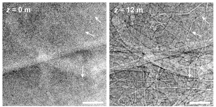
They reported that phase-contrast chest radiography can visualize the smallest airways—measuring less than 2mm—and their disease-related obstructions. The study’s lead authors, Ilian Häggmark and Kian Shaker, researchers at KTH Royal Institute of Technology, say that these are details that don’t show up in conventional radiography.
The researchers reported their findings in the Proceedings of the National Academy of Sciences (PNAS) of the United States of America.
Phase contrast imaging is used in research labs with equipment that today is limited in use to imaging centimeter-scale samples of soft tissue. But, Häggmark says, the study clearly shows that it’s possible to do more with phase-contrast X-ray imaging, if the technical demands for clinical use can be engineered.
The chest radiography that clinics and hospitals use today plays an important role in detecting respiratory disease, but it is fundamentally limited by the way in which it generates images, Häggmark says.
He says that the promising phase-contrast technique used in the study could show subtle pathological changes that are otherwise invisible with conventional X-ray imaging, which is important when screening for diseases like asthma or chronic obstructive pulmonary disease (COPD).
“Phase-contrast X-ray imaging can extract more information at higher resolution using the same amount of radiation dose as in conventional radiography,” Häggmark says.
In conventional radiography, the X-ray beam passes through the body, where it is absorbed along the way in different tissues by different amounts. On the other side, a detector measures the intensity of the beam—or what’s left of it—after it has been filtered through the body. This process is known as attenuation, and it’s the basic mechanism for providing the contrast that makes X-ray images useful.
The phase-contrast technique is a way of getting more information out of each X-ray beam. That’s because it’s possible to measure differences in the waveforms of X-rays that pass through a sample. X-ray beams encounter atoms and other structures that can change the position of the wave at any point in time—the phase—in relation to a reference wave. This phase information is used to generate an image that enhances structures in the sample, which in the human chest highlights the boundaries of bronchial walls and small airways with higher contrast and better resolution.
Häggmark says that one key to the method is to move the detector further away from the patient.
Development of equipment for imaging larger samples will take time, he says. “You need an X-ray source with both high power and a small emission spot,” he says. “Basically you need bright X-ray sources.”
He says that promising developments are being carried out, but it will take time for this to reach testing for human use,” he says.
“For now, simulations and virtual clinical trials are the perfect tools to explore what we can do when the source technology is ready.”
Keywords
Images
About KTH Royal Institute of Technology

Founded in 1827, KTH Royal Institute of Technology in Stockholm is one of the world's leading technical and engineering universities, as well as a key center of intellectual talent and innovation.
Subscribe to releases from KTH Royal Institute of Technology
Subscribe to all the latest releases from KTH Royal Institute of Technology by registering your e-mail address below. You can unsubscribe at any time.
Latest releases from KTH Royal Institute of Technology
New chip offers way to make use of quantum system ‘imperfections’19.5.2026 14:30:22 CEST | Press Release
Quantum technologies promise powerful new kinds of computers, giving scientists new tools to mimic and explore nature at its tiniest scales. At those levels, everything in nature—from atoms and electrons to light itself—follows the strange rules of quantum mechanics. But the real world is never perfectly clean: signals fade, energy leaks away and systems pick up noise from their surroundings.
Twisting atom thin materials reveals new way to save computing energy6.5.2026 13:06:14 CEST | Press Release
A recent study shows a new and potentially more energy efficient way for information to be transmitted inside electronic systems, including computers and phones—without relying on electric currents or external magnetic fields.
Cause of common heart valve defect revealed in genetic study28.4.2026 11:18:02 CEST | Press Release
New clues from genetic research may help explain what causes the most common heart defect present at birth. Researchers in Sweden have identified rare DNA changes during fetal development that can lead to a condition known as bicuspid aortic valve (BAV).
Study offers new way to stop global potato pathogen once linked to Ireland’s Great Famine23.4.2026 11:46:21 CEST | Press Release
Scientists in Sweden have taken an important step toward fighting potato late blight, a plant disease that once triggered an historic famine in Ireland and now threatens to spread globally due to climate change.
Why do incomplete spinal cord injuries cause unstable movements? New study reveals disrupted muscle coordination14.4.2026 12:09:09 CEST | Press Release
Even when people with incomplete spinal cord injuries can walk, everyday functions like standing, balancing or producing steady force may remain difficult. A new study shows why.
In our pressroom you can read all our latest releases, find our press contacts, images, documents and other relevant information about us.
Visit our pressroom